
What Is TLIF? Understanding L4-L5 Transforaminal Lumbar Interbody Fusion
By Joey Montes · September 19, 2026
TLIF stands for Transforaminal Lumbar Interbody Fusion. It is a spinal fusion procedure designed to stabilize a segment of the lumbar spine while also allowing the surgeon to reach the disc space and nearby nerve structures.
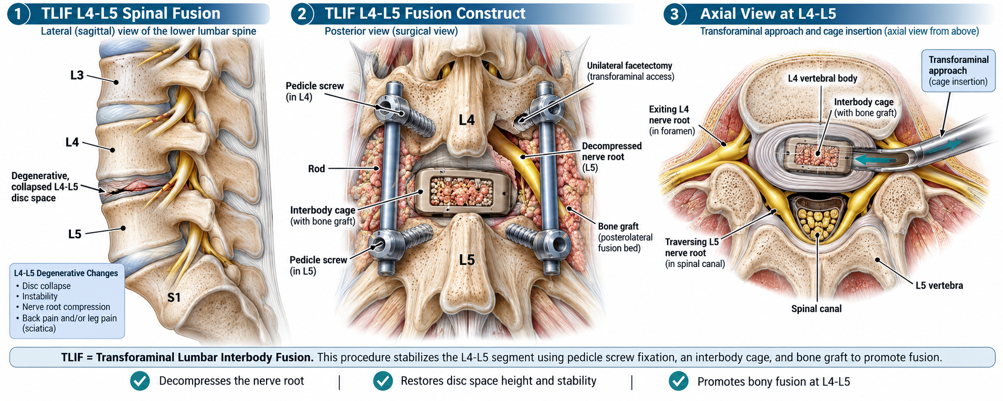
The illustration above focuses on a TLIF performed at L4-L5, the level between the fourth and fifth lumbar vertebrae. It presents the procedure from three perspectives: a side view of the lumbar spine, a posterior view of the completed fusion construct, and an axial view showing the transforaminal approach to the disc space.
What happens at L4-L5?
The first panel shows the lower lumbar spine from the side. The L3, L4, L5, and S1 levels are identified, with attention centered on the L4-L5 disc space.
In the illustration, the L4-L5 disc space is shown as degenerative and collapsed. The graphic associates these changes with several possible problems:
- Disc collapse
- Instability
- Nerve root compression
- Back pain and leg pain, including sciatica
The goal of a fusion procedure is to stabilize the affected spinal segment and create an environment in which L4 and L5 can eventually form a solid bony fusion.
What does TLIF mean?
The name describes both the location and the surgical approach:
- Transforaminal refers to approaching the disc space from one side through the region near the neural foramen.
- Lumbar refers to the lower portion of the spine.
- Interbody refers to the space between two vertebral bodies.
- Fusion refers to encouraging the treated vertebrae to grow together into a stable unit.
At L4-L5, the procedure is performed between the L4 and L5 vertebrae.
The posterior fusion construct
The second panel shows the spine from the back after the fusion construct has been placed.
Several important parts of the procedure are visible.
Pedicle screws
Pedicle screws are shown in both L4 and L5. These screws provide strong fixation points within the vertebrae.
Rods
Metal rods connect the screws on each side. Together, the screws and rods provide mechanical stability to the L4-L5 segment while the biological fusion develops.
Interbody cage
An interbody cage is positioned between L4 and L5. In the illustration, the cage contains bone graft.
The cage occupies the disc space and helps support the space between the two vertebrae while fusion takes place.
Bone graft
The illustration also shows bone graft placed around the posterior portion of the fusion construct. Bone graft provides material intended to support new bone growth and the development of a solid fusion.
Nerve decompression
The posterior view labels a decompressed L5 nerve root and also shows a unilateral facetectomy used to provide transforaminal access.
This part of the operation creates access to the disc space while also providing room around the affected nerve structures.
The transforaminal approach
The third panel provides an axial view, looking down at L4-L5 from above.
This perspective helps explain why the operation is called transforaminal. The surgical instruments approach the disc space from one side rather than directly through the center of the spinal canal.
The illustration identifies several important structures:
- The L4 vertebral body
- The L5 vertebra
- The spinal canal
- The exiting L4 nerve root
- The traversing L5 nerve root
- The interbody cage and bone graft
The cage is inserted into the prepared disc space through this transforaminal pathway.
What is the interbody cage for?
The interbody cage is one of the central components of a TLIF.
It is positioned between the vertebral bodies after the disc space has been prepared. In the illustration, the cage is packed with bone graft.
Its role is to support the L4-L5 segment while providing an environment in which bone can grow through and around the treated area.
The graphic specifically highlights three intended results of the procedure:
- Decompression of the nerve root
- Restoration of disc-space height and stability
- Promotion of bony fusion at L4-L5
Why are screws and rods needed?
The screws and rods provide immediate structural support to the treated spinal segment.
The hardware itself is not the biological fusion. Instead, it stabilizes L4 and L5 while bone growth develops over time.
This distinction is important. The surgical construct can be placed during the operation, but the process of forming a mature bony fusion takes much longer.
What does decompression mean?
Decompression refers to creating more space around a nerve that is being crowded or compressed.
In this illustration, the L5 nerve root is specifically identified as decompressed. The axial view also shows the relationship between the exiting L4 nerve root, the traversing L5 nerve root, and the surgical pathway.
That relationship is one of the reasons careful surgical planning is important during a TLIF.
TLIF combines stabilization and fusion
A useful way to understand TLIF is to think of it as several related goals being addressed during one operation.
The procedure can involve removing damaged disc material, creating space around affected nerve structures, placing an interbody cage, adding bone graft, and stabilizing the vertebrae with screws and rods.
A TLIF is not simply the placement of spinal hardware. The long-term objective is a stable bony fusion between the treated vertebrae.
What happens after surgery?
The illustration explains the surgical anatomy, but it does not represent an individual recovery timeline.
After a TLIF, the body needs time to heal and for fusion to develop. The screws, rods, cage, and bone graft create the structural environment for that process, but bone growth occurs gradually.
Activity restrictions, walking, rehabilitation, follow-up examinations, and imaging depend on the individual patient and the treating surgical team.
The L4-L5 TLIF in one view
The three panels of this illustration provide a useful summary of the procedure.
The first shows the problem: a degenerative and collapsed L4-L5 disc space.
The second shows the reconstruction: pedicle screws, rods, an interbody cage, bone graft, and decompression of the nerve root.
The third shows the surgical pathway: the transforaminal route used to reach the disc space while working around the nearby L4 and L5 nerve roots.
Together, they show why TLIF is both a decompression and stabilization procedure, with the ultimate goal of promoting a solid fusion at the treated lumbar level.
In summary
TLIF stands for Transforaminal Lumbar Interbody Fusion.
At L4-L5, the procedure uses an interbody cage, bone graft, pedicle screws, and rods to stabilize the segment and promote fusion. The transforaminal approach gives the surgeon access to the disc space from one side, while decompression can create more room around affected nerve structures.
The hardware supplies stability, while the bone graft supports the longer biological process of fusion.
This article is an educational overview based on the illustrated L4-L5 TLIF construct shown above. The exact procedure, surgical technique, indications, restrictions, and recovery plan vary by patient and should be discussed with the treating spine surgeon.